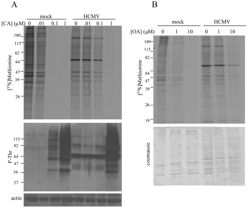
Figure 3.
Protein synthetic activity and protein phosphorylation in HCMV-infected cells treated with CA and okadaic acid (OA). (A) Mock-infected or HCMV-infected HFs (72 hpi) were treated with increasing concentrations of CA for 30 minutes followed by [35S]methionine ([100 µCi/ml] labeling for 30 minutes. De novo protein synthesis was assessed by SDS-PAGE and autoradiography (top panel) and the pattern of protein phosphorylation was determined by immunoblot analysis using a phospho-threonine (P-Thr) specific antibody (middle panel). Equivalent protein loading was confirmed by immunoblot analysis for actin (bottom panel). (B) Mock-infected HFs or HCMV-infected HFs were treated with the indicated concentration of OA for two hours beginning at 72 hpi. De novo protein synthesis (top panel) was assessed as described above after Coomassie G-250 staining (bottom panel) to document equivalent protein loading.

