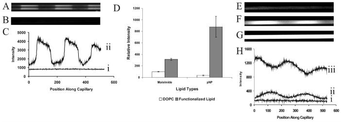
Figure 5.
Covalent functionalization of poly(lipid) patterned capillaries. A) Fluorescence image of a poly(bis-SorbPC) patterned capillary in which the void regions were filled with maleimide-doped DOPC SUVs, followed by covalent immobilization of FQ-labeled, reduced ribonuclease A. Scale bar = 60 μm. B) Fluorescence image of poly(bis-SorbPC) patterned capillary with maleimide-doped DOPC, followed by introduction of FQ-labeled unreduced ribonuclease A. C) Linescans showing the fluorescence intensity of capillaries shown in Figure 5A (ii) and Figure 5B (i) as a function of position along the capillary (pixel number). The linescans were taken along the inner capillary wall. D) Relative fluorescence intensity versus functionalization (± 1 s.d.), n=3 for each capillary type. Bare regions of the pattern were functionalized using DOPC (white bars) or DOPC doped with headgroup functionalized lipid (gray bars). E) Fluorescent image of poly(bis-SorbPC) patterned capillary with the void regions filled with DOPC following introduction of β-galactosidase and fluorescein di( β-D) galactopyranoside (FDG) (t = 3 min). F) Fluorescent image of poly(bis-SorbPC) patterned capillary containing β-galactosidase immobilized via 20% maleimide-doped DOPC following introduction of FDG (t = 30 s). G Fluorescent image of poly(bis-SorbPC) patterned capillary containing β-galactosidase immobilized via 20% maleimide-doped DOPC following introduction of FDG (t = 3 min). Images in E, F and G are shown on the same scale. G) Linescans through the center of capillaries shown in Figure 5E (i), Figure 5F (ii), and Figure 5G (iii).

