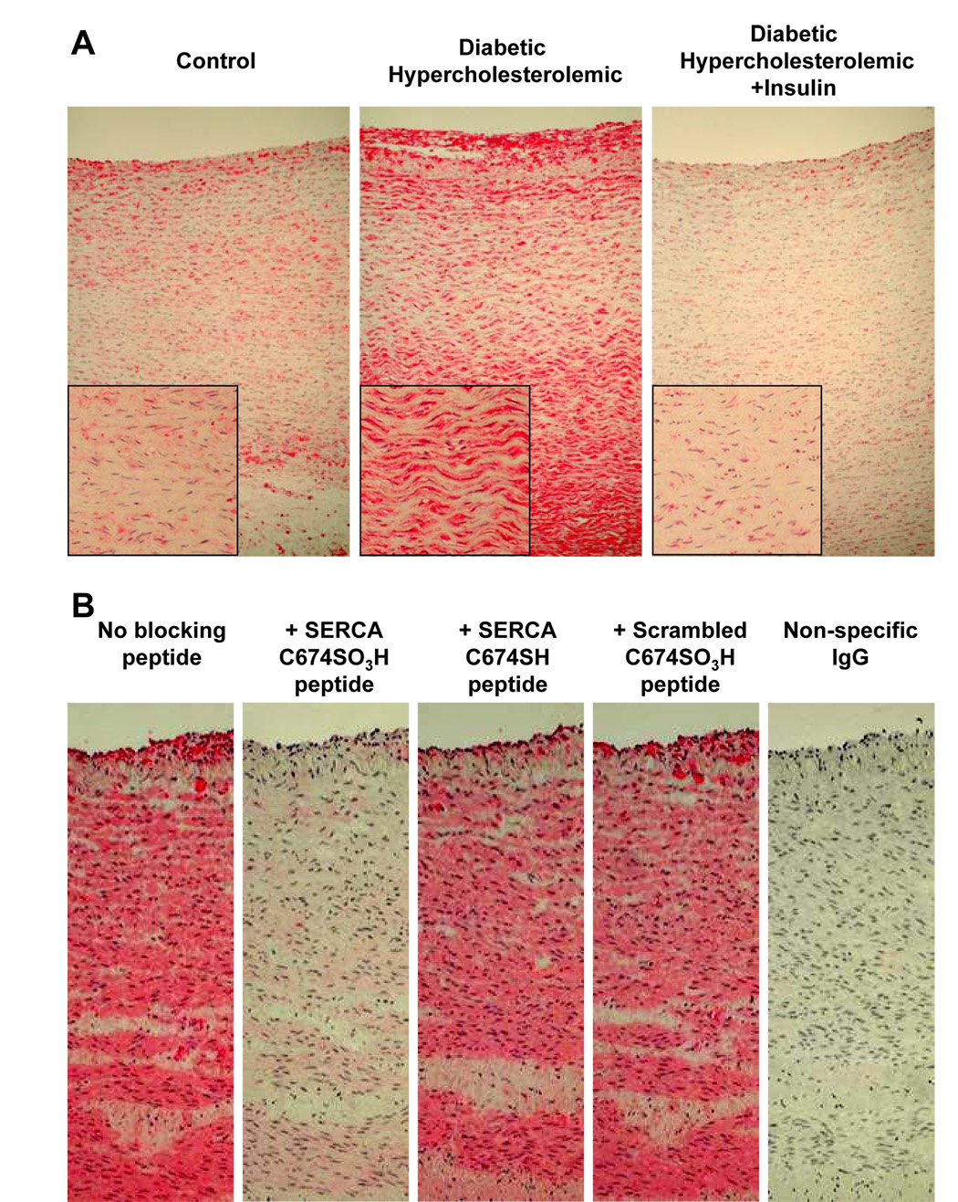
Figure 2. Immuno-histochemical staining with anti-SERCA C674-SO3H of aorta from control pig (#1), diabetic hyperlipidemic pig (#4) and diabetic pig treated with insulin (#7).
A: Positive staining is shown in diabetic hyperlipidemic pig (#4) with less intense staining observed in other pigs (see Supplemental Figure 2B). Staining can be seen in both the plaque on the lumenal aspect of the diabetic pig aorta as well as in the smooth muscle lamellae.
B: Specificity of staining with anti-SERCA C674-SO3H antibody in aorta of diabetic hyperlipidemic pig #6. Staining was blocked by the C674-SO3H SERCA peptide, but not by the SERCA peptide with reduced cysteine-674, or the scrambled C674-SO3H SERCA peptide, indicating specific staining of SERCA C674-SO3H. Anti-rabbit IgG secondary antibody staining was negative.

