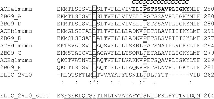
Figure 1.
Alignment of amino acid sequences spanning the M2 segment, the M2M3 loop, and part of the M3 segment of the mouse muscle nAChR α (ACHa1mumu), β (ACHb1mumu), γ (ACHg1mumu), and δ (ACHd1mumu) subunits, and Torpedo nAChR α (2BG9_A, 2BG9_D), β (2BG9_B), γ (2BG9_E), and δ (2BG9_C) subunits (PDB entry 2BG9). Segments that are α-helical in the Torpedo nAChR structure are underlined. The conserved 9′ leucine and the conserved 23′ proline are boxed. The 16 positions in the α-subunit investigated in this study that were individually mutated to Cys are in bold and also indicated by a bold italic “C” above the residue. Two possible alignments for the prokaryotic Cys-loop receptor homolog from ELIC are shown. The upper alignment was obtained by aligning all known Cys-loop receptor sequences; the lower alignment is a structure-based alignment from Hilf and Dutzler (2009). Numbers at the right of each row indicate the amino acid number of the last residue shown. Symbols below alignment denoting the degree of conservation observed in each column are as follows: an asterisk means that the residues or nucleotides in that column are identical in all sequences in the alignment, a colon means that conserved substitutions have been observed, and a period means that semiconserved substitutions are observed.

