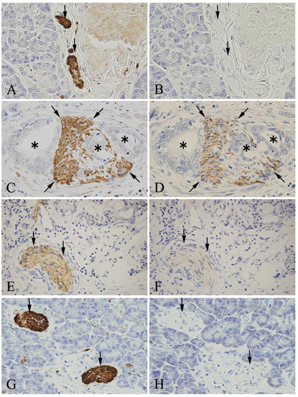
Figure 7. Immunohistochemical analysis of Nestin in nerve fibers.
Peripheral nerve fibers were stained with anti-S-100 protein (A, C, E and G, arrows). Nestin was not present in the nerve fibers in normal pancreatic tissues (B, arrows), but was present in nerve fibers (D, arrows) invaded by cancer cells (D, asterisks). Nestin was also present in the nerve fibers in chronic pancreatitis (CP)-like lesions that were adjacent to the cancer cells (F, arrows), but not when such lesions were distant from the cancer cells (H, arrows).
Original magnification: A-H×400

