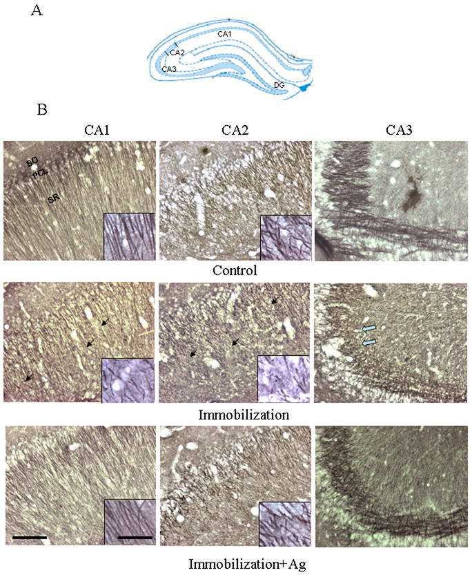
Figure 2.

A. Schematic representation of the hippocampal subfields in the coronal plane (approximately Bregma -3.24 mm) (Paxinos and Watson 2005). B. Photomicrographs showing the CA1, CA2 and CA3 of the rat hippocampus immunostained with β-tubulin III after the injection of saline (Control), repeated immobilization plus saline (Immobilization) and repeated immobilization plus agmatine (50 mg/kg/day, i.p.) (Immobilization+Ag) for seven days (n=7 for each group). PCL, pyramidal cell layer; SO, stratum oriens; SR, stratum radiatum. Scale bars = 100 μm for the pictures except for inserts; 20 μm for inserts. Solid arrows point to areas where showed less intensity of immunostaining in the CA1 and CA2 of stress animals, compared to those of the control and immobilization plus agmatine. The open arrow points to an area in the CA3 with reduced mossy fibers in stress animals.
