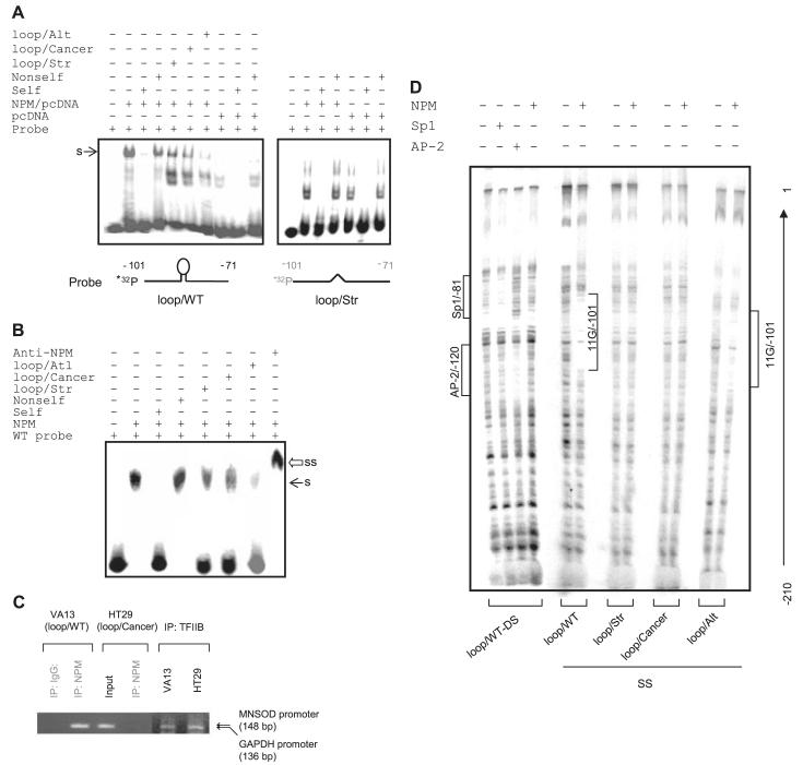
FIGURE 4. Interaction of NPM with the 11G-loop in the human sod2 promoter region.
A, nuclear extracts from NPM-transfected cells or vector-transfected cells (control) were probed using single-stranded probe: loop/WT (left) or loop/Str (right). B, NPM protein was incubated with the loop/WT probe. 100-fold self, non-self, loop/Str, and loop/Cancer competitors were added to identify the specific binding (s) indicated by arrows (A and B). A supershift band (ss) by NPM antibody is indicated by an open arrow (B). C, chromatin was precipitated from VA13 cells with the wild-type sequence (right) or HT29 cells with cancer-type sequence (left) using NPM or TFIIB antibody. The sod2 and GAPDH promoter fragments (indicated by arrows) were detected by PCR. D, DNase I footprinting analysis by incubating 32P-labeled double- or single-stranded promoter regions with NPM, Sp1, and AP-2 proteins and followed by DNase I digestion. Protected regions are indicated for Sp1/AP-2 binding motif and the 11G sequence bound with NPM.

