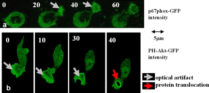
Figure 7.
Fluorescence intensity increases of GFP constructs in forming pseudopodia. The artifactual increases in fluorescent signal are evident in forming pseudopodia in neutrophilic HL60 cells expressing p67phox-GFP (a) and PH-Akt-GFP (b). The optical artifact is evident in the forming pseudopodia of these cells (gray arrows). However, this artifact can be distinguished from the genuine translocation of PH-Akt-GFP to the phagosomal membrane (red arrow). Bars, 5 µm

