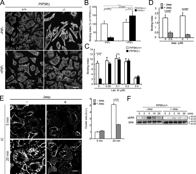
Figure 3.
The relation between PIP5K-γ deficiency, actin, particle attachment, and FcγR clustering defects. (A) Rescue of cell shape and actin by PIP2 shuttling. BMM with or without exogenously added PIP2 were stained with phalloidin. (B) Rescue of particle attachment by PIP2 delivery. Binding indices (n > 100) are expressed as the percentage of WT BMM without PIP2. (C) Rescue of particle attachment by Latr B. Cells were incubated with Latr B for 5 min at 37°C before incubation with particles at 4°C (n > 70). (D) Inhibition of particle attachment to WT BMM by Jasp. Cells were incubated with 1 µM Jasp at 37°C for 30 min before the addition of IgG beads (n > 50). (E) Inhibition of FcγR clustering in WT BMM by Jasp. (left) Fluorescence staining of FcγR–IC clusters. (right) Cluster size quantitation (n > 1,000). Error bars indicate SEM. (F) Attenuation of IC-induced ERK phosphorylation by Jasp. Cells pretreated with or without Jasp were incubated with IC at 4°C for 20 min and warmed to 37°C. Bars, 10 µm.

