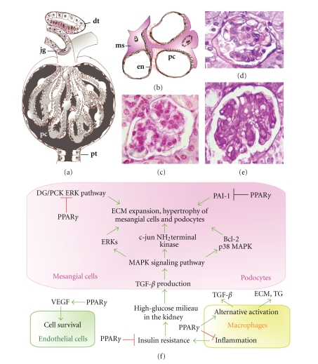
Figure 1.
Roles of PPARγ in the filtration units of the kidney. The kidney capsules (a) contain the glomerular capillaries covered with podocytes (pc). In the wall of the afferent arterioles, modified smooth muscle cells form the juxtaglomerular system (jg). The filtrated urine is guided to the proximal tubules (pt). The distal tubules (dt) can return to the cortical kidney capsules and their epithelial layers serve as a chemosensory region, the macula densa (labeled with red). (b) PPARγ activation affects either podocyte (pc), mesangial cell (ms), or endothel cell (en) functions. (c) Periodic acid-Schiff (PAS) stained sections of a normal kidney capsule in mouse. (d) Glomerulonephritis in high-fat diet fed mouse and (e) type 2 diabetic (db/db) mouse, showing intensive PAS staining of the expanded mesangial matrix, thickening of glomerular walls, and enlargement of kidney capsules. (f) Summary of PPARγ-mediated cellular events in mesangial cells, podocytes, kidney macrophages, and glomerular endothel cells.

