Figure 3.

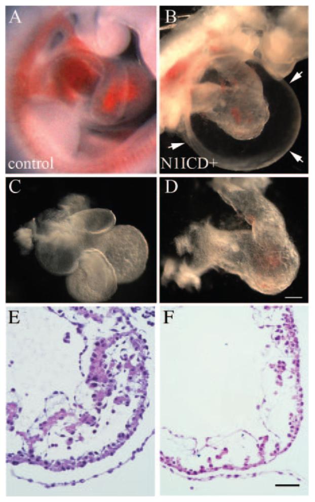
Heart defects in Notch1ICD+ embryos. Freshly dissected E9.5 wild-type (A) or N1ICD+ (B) embryos are shown. Isolated hearts from N1ICD+ embryos displayed abnormal ventricular looping and underdeveloped cardiac chambers (D), compared to the normally developed heart (C). The scale bar in D represents 100 μm for A and B, 70 μm for C, and 60 μm for D. E and F, Heart sections from control (E) or N1ICD+ (F) embryos were hematoxylin/eosin-stained to show ventricular trabeculation. The scale bar in F represents 25 μm for E and F.
