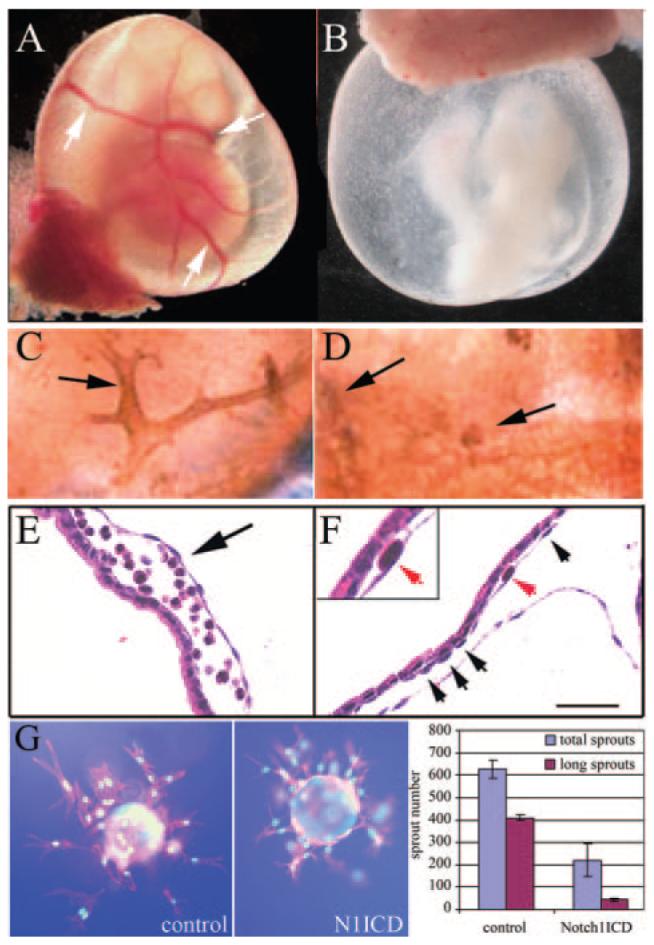
Figure 4.

Defects in yolk sac vasculature in Notch1ICD+ embryos. Embryos were collected from control (A, C, and E) or N1ICD+ embryos (B, D, and F) at E9.5. Whole mount views of embryos with intact yolk sacs show lack of conducting arteries (arrows, A) and orange peel—like appearance of N1ICD+ yolk sacs (B). C and D, PECAM-1-staining shows normal vascular structure (C) vs the fused primitive vascular network in N1ICD+ yolk sacs (D). E and F, hematoxylin/eosin-stained sections contrast normal blood filled vessels in control (E) with a gross enlargement between endoderm and mesoderm layers in N1ICD+ embryos (F), resulting in lacunae-like spaces. Endothelial cells were present in the N1ICD+ yolk sacs (F, arrows, inset). The scale bar in F represents 50 μm. G, Notch1ICD inhibits endothelial sprouting in vitro. Fibronectin-coated microcarrier beads were seeded with HUVEC-GFP or HUVEC-Notch1ICD, embedded in fibrin, and grown for 3 days. Normal branching was inhibited by Notch1ICD. Total sprouts and sprouts > 150 μm (long sprouts) were counted and measured and shown as means± SD.
