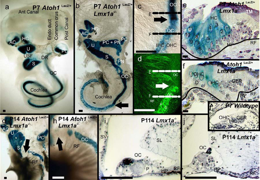
Fig. 3. Postnatal Lmx1a mutant ears reveal disorganized sensory epithelia.

A. In the wildtype, six discrete sensory epithelia are separated from one another by constricted, non-sensory epithelial spacers. Two of three semicircular canals and an endolymphatic duct can be identified as shadows. B. In the Lmx1a mutant, the anterior and horizontal cristae are separated by a common cruciate eminence, while the posterior crista is grossly enlarged and extended by the presence of both embedded and detached papilla neglecta-like sensory epithelia (PN). The utricle, saccule and cochlear sensory epithelia appear continuous with one another. The basal turn of the organ of Corti appears as a uniform band of hair cells that is discretely separated (arrow) from an apex in which inner and outer hair cells can be identified. C, Higher magnification view of the arrowed transition in B. The densely packed hair cells of the basal cochlea are above the arrow and the apex below it. The black lines indicate the planes of section in E and F. D. Same tissue as C, but stained for beta-tubulin to reveal nerve fibers and pillar and Deiter’s cells. Note the absence of tubulin-containing pillar and Deiter’s cells in the base and their conspicuous appearance at the transition to the apex. E. A medio-lateral near radial section across the base of the mutant cochlear duct as indicated by the dotted line in C. Note the presence of a tectorial membrane (TM). Up to 11 rows of hair cells are marked by the blue Atoh1LacZ reaction product, unlabelled supporting cells are present below the hair cells. F. A recognizable organ of Corti with inner and outer hair cells is present in the apex (compare with wildtype insert). G,H Beginning around P14, hair cells disappear, starting in the base (arrow). In contrast, nerve fibers continue to mature as indicated by the osmium tetroxide stained myelin (black fibers). I., J. By P114 the organ of Corti is grossly disorganized, lacks identifiable hair cells and shows massive aberrations in almost all associated epithelia such as the spiral limbus (SL) and the stria vascularis (SV). These data suggest that absence of Lmx1a is ultimately incompatible with hair cell maintenance though that its absence does not interfere with their initial formation. Scale bars are 100µm.
