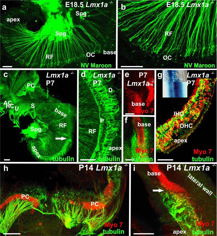
Fig. 4. Late innervation and sensory epithelia are disorganized.

A, B Afferent radial fibers to the base of the E18.5 mutant cochlea stained with the lipophylic dye, NV Maroon (A,B) and anti-acetylated tubulin (C,D,F-I). Note that the fibers enter the organ of Corti but do not extend to the outer hair cells as they would in a comparable wildtype ear. There is a notable difference between the packing density of radial fibers in the base and the apex consistent with the reduced presence of spiral ganglia in the base. C There is a complete absence of pillar/Deiter’s cells basal to the cochlear transition (arrow) and clear distinction of innervations between the densely innervated saccule and the poorly innervated basal turn of the cochlea. D. Disorganized Deiter’s cell processes (D) in the apex of the ear in A (now stained for β-tubulin) show longitudinal extension along the cochlea. E. Myo7a staining shows close proximity of OC and papilla neglecta hair cells in the basal cochlea of a P7 mutant. F. The basal/apical cochlear transition of the ear in C., with β-tubulin stained supporting cells and Myo7a stained hair cells. Note that the packing density of hair cells is inversely related to supporting cell labeling. This pattern is maintained as long as hair cells can be labeled by Myo7a antibodies (I). G. Apical cochlea of the ear in C/D. IHC’s, and OHC’s can be recognized, but the organization is inferior to that of the wildtype (see Atoh1lacZ stained hair cells in the inset, mutant apex above, wildtype below). H. The grossly enlarged and elongated posterior crista of a P14 mutant shows fibers targeted to the Myo7a positive hair cells. Scale bars are 100 µm.
