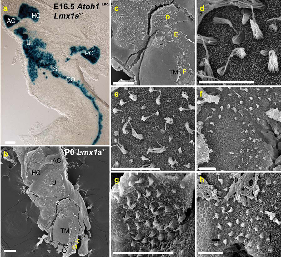
Fig. 6. Continuity of hair cells in late embryos as revealed by Atoh1LacZ staining and SEM.

A The sensory epithelia of the E16.5 Lmx1a mutant ear revealed by Atoh1lacZ staining of hair cells. Note that the juxtaposition of sensory epithelia found at P7 in Fig. 1B is already apparent at E16.5. B. A P0 Lmx1a mutant ear oriented similar to that in A and viewed in the scanning electron microscope. The positions of the micrographs in G and H are indicated at the bottom of the micrograph. C. A higher power micrograph centered on the “S” (saccule) of B. The positions of micrographs in D–F are indicated, as is the smooth, flat surface of the tectorial membrane (TM). D. Micrograph of the saccular macula. The sterocilia come in long and immature short variations of the pipe-organ arrangement characteristic of vestibular hair cells. E. A region of the basal cochlea (note the adjacent tectorial membrane in C) close to the saccule. A mix of hair cells with long, vestibular-like and short, C-shaped cochlear inner hair cell-like sterocilia are present. F. Further toward the apex, but still in the basal cochlea, vestibular type hair cells are found adjacent to the tectorial membrane, and cochear-like hair cells further lateral. G. Still in the base, but further toward the apex, multiple “rows” of hair cells all possess short, cochlea-like sterocilia bundles. H. Adjacent to G. in the base, variously oriented cochlear hair cells are present (arrows indicate polarity). For abbreviations, see Fig.1. Medial is left and lateral right in Fig’s. 3 D–H. Scale bars are 100µm.
