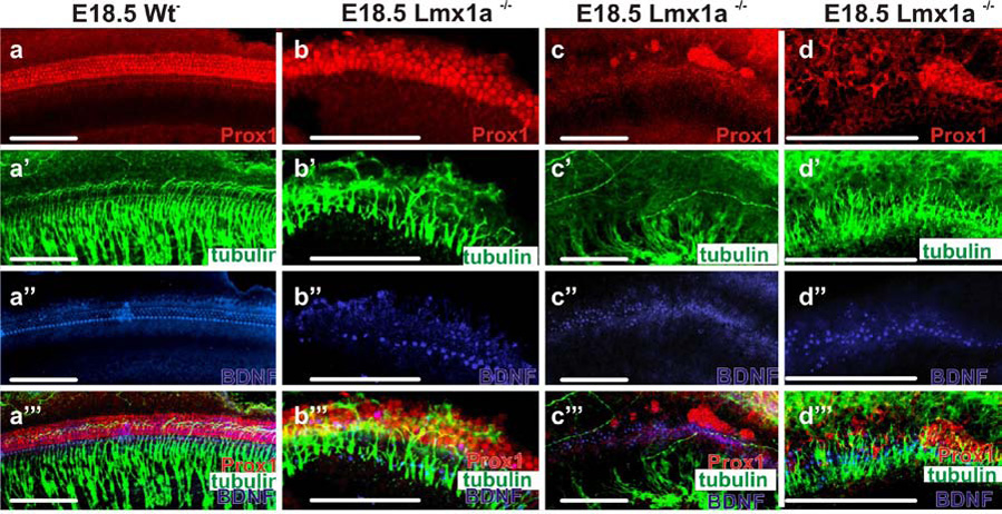
Fig. 7. Hair cells, supporting cells and nerve fibers are disorganized.

E18.5 organs of Corti immunohistochemically (ImHC) stained for Prox1, β-tubulin, BDNF, and all three combined (rows A-A’’’), in the wildtype, mutant apex, mutant base, and high mag. mutant base (columns A–D). A–D Prox1 ImHC. there is an orderly expression in supporting cells in wildtype, some organization in the apex in the mutant but clumping of PROX1 stained supporting cells in the mutant base (see also A’’’–D’’’). A’–D’ β-tubulin ImHC. Note that nerve fibers in the base stop short of the OC (see also A’’’ – D’’’). A’’–D’’’ BDNF ImHC stains IHC’s strongly and OHC’s weakly in the wildtype. A row of IHC’s can be recognized in the mutant apex, but strongly stained cells are scattered in the base. A’’’ – D’’’ The superimposed images show that hair cells and supporting cells overlap and orderly nerve fibers project between the supporting cells in wildtype whereas hair cells and supporting cells do not overlap in mutants with disorganized fibers projecting between the labeled supporting cells (apex) or reaching hair cells (base) Scale bars are 100µm.
