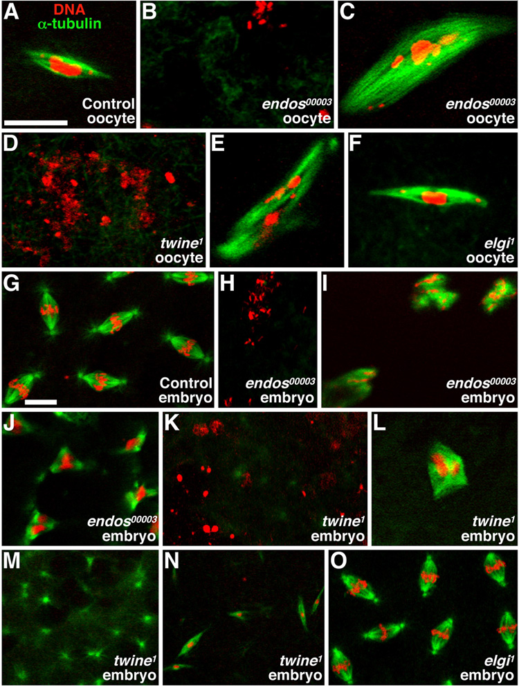
Figure 2. endos00003 mutants have spindle defects.
Stage 14 oocytes (A–F) and 0–3 hour embryos (G–O) stained with propidium iodide (DNA, red) and anti-α-tubulin (microtubules, green). (A) Control oocytes in metaphase I. (B) Typical endos00003 oocyte showing dispersed DNA without a spindle. (C) Rare endos00003 oocyte showing abnormal DNA associated with large spindle masses. (D,E) Similar twine1 phenotypes. (F) elgi1 oocyte in metaphase I. (G) Control embryos exhibiting typical embryonic mitoses. (H–J) endos00003-derived embryos showing dispersed DNA with no spindle (H), dispersed DNA with spindle masses (I), and tripolar spindles (J). twine1-derived embryos showing dispersed DNA (K), abnormal spindle masses (L), spindle asters with no DNA (M), and long, thin spindles (N). (O) Embryos derived from elgi1 females showing normal spindles and DNA. Scale bars, 10 µm.

