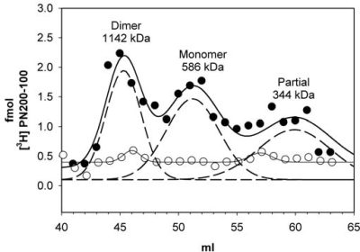
Fig. 4.
Isolation of native CaV1.2 as mixtures of monomers and dimers. Cardiac microsomes were labeled with 20 nM [3H]PN200–110, solubilized with 1% digitonin and proteins were separated on a Superdex 200 column. Total bound [3H]PN200–110 is plotted for each 1-mL fraction in the absence (filled circle) or presence of isradipine (open circle). Gaussian fit for the data are plotted as a solid line and the individual Gaussians are plotted as dashed lines.

