
FIGURE 3.

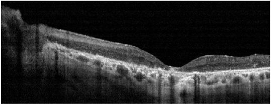

Case 1 (KL)
Fundus photograph of patient KL shows a bullseye maculopathy with surrounding bone spicules (Top left). Horizontal OCT scan shows corresponding hyperreflectivity of the choroidal layer with overlying marked retinal and RPE thinning (Top right) The corresponding retinal thickness profile of KL (solid line) compared with normal controls (mean with 95 % CI shown as grey band) (Bottom).