FIGURE 4.
case 2 JW RP 20/20 OU
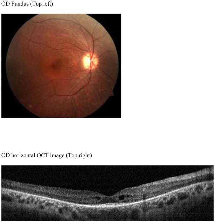
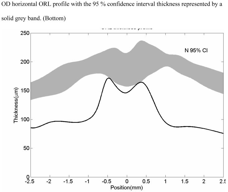
Patient JW, early RP. Color fundus photo shows a mild macular bullseye lesion with arterial attenuation but no bone spicules (Top left). Horizontal OCT scan shows macular thinning and perifoveal cysts (Top right). Horizontal ORL profile shows that the mean values of this patient with early RP is below the normal average (normal 95 % confidence interval thickness represented by a solid grey band.) (Bottom).


