
FIGURE 5.

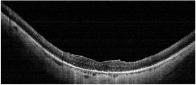

Case 4 DK, Cone-rod, 20/200 no choriocapillaris
Patient DK. Color fundus photo shows macular atrophy, vascular attenuation and absence of RPE pigment spicules (Top left). Horizontal OCT scan shows macular thinning and marked loss of the choriocapillaris. An overlying epiretinal membrane is visible (Top right). Horizontal ORL profile shows marked thinning of the macular area as compared with normals (solid grey band) (Bottom).