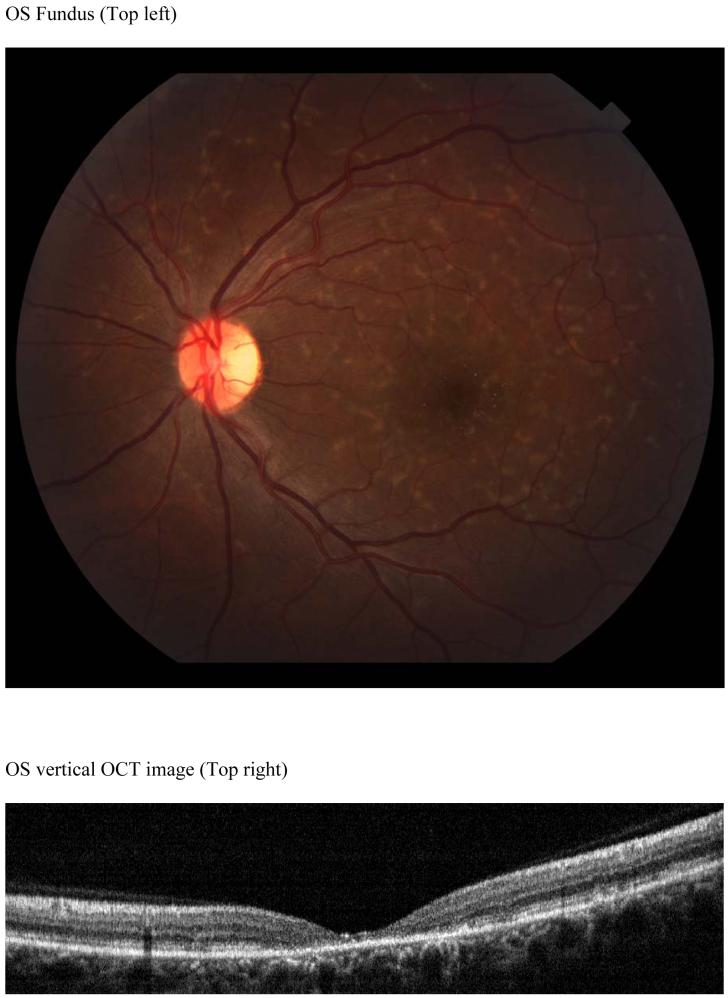
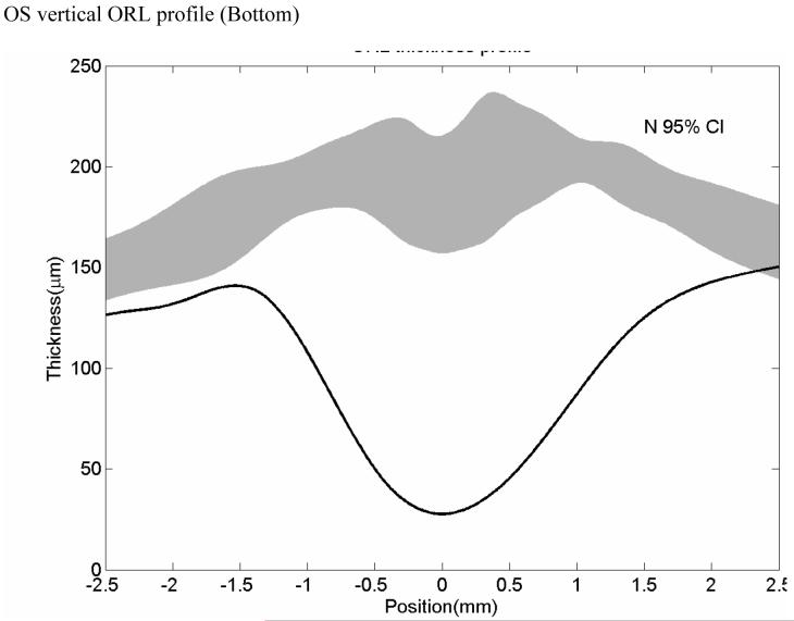
FIGURE 6.
Case 6 MM Stargardt, 20/ 70
MM (Stargardt) Color fundus photo shows numerous pisciform yellow flecks, normal appearing RPE and absence of atrophy (Top left). Horizontal OCT scan reveals a thinned perifoveal retina with normal adjacent retinal thickness (Top right). Retinal thickness profile shows this marked retinal thinning centrally (deep central dip) as compared with normal controls (grey band) (Bottom).


