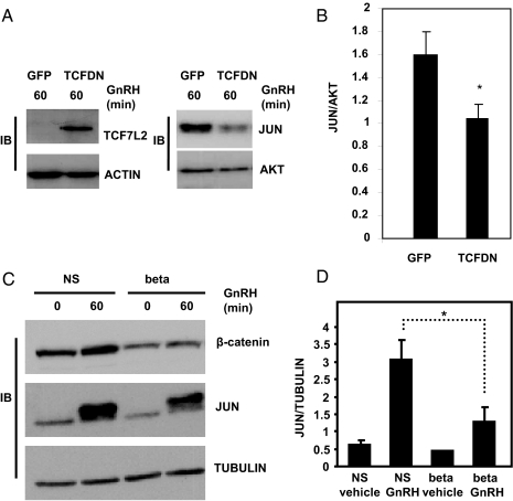
Figure 4.
JUN protein levels are reduced in β-catenin-deficient and TCFDN-expressing cells. A, Whole-cell extracts isolated from LβT2 cells transduced with either GFP- or TCFDN-expressing adenoviruses (5 × 10¹⁰ vpm) and treated with 100 nm GnRH for 1 h, were subject to immunoblot analysis using antibodies specific for TCF7L2, JUN, actin (loading control), and AKT (loading control). B, Arbitrary densitometric units of JUN divided by arbitrary densitometric units of AKT. Data shown in B are the means ± sem from three distinct experiments. *, P < 0.05. C, Whole-cell extracts were isolated from LβT2 cells stably expressing nonsilencing shRNA (NS) or shRNA to β-catenin (beta) and subjected to immunoblot analysis using antibodies specific for β-catenin, JUN, and tubulin (loading control). D, Arbitrary densitometric units of JUN divided by arbitrary densitometric units of tubulin. Data shown in D are the means ± sem from three distinct experiments. *, P < 0.05.

