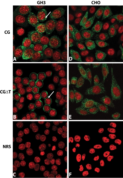
Figure 5.
Immunofluorescent localization of CG or CGΔT dimers in GH3 (A–C) and CHO (D–F) cells. The cells were immunostained with CGβ antiserum (A, B, D, and E) or normal rabbit serum (NRS; C and F represent CGΔT cells), and immunofluorescence was detected by confocal microscopy (green, positive staining with the primary antiserum; red, nuclear staining). Note the unique honeycomb pattern (arrow) of staining for CGΔT (B) vs. dispersed puncta (arrow) for CG dimer (A). There was no difference in the staining pattern for CG (D) and CGΔT (E) in CHO cells with the lattice staining pattern of CGΔT conspicuously absent in these cells. The micrographs shown are representative of eight experiments and are at ×60 magnification.

