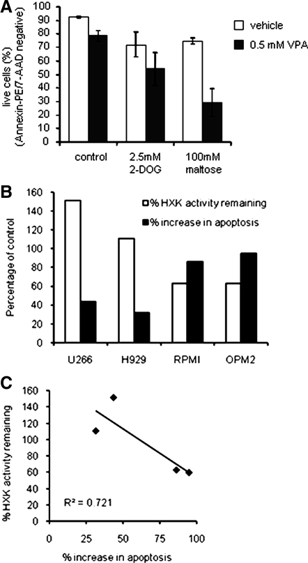
Figure 5.
HDACI cooperates with other glycolytic inhibitors to induce apoptosis. A, OPM2 cells were treated with vehicle, 2-DOG, or maltose for 72 h in the presence or absence of VPA. Apoptosis was analyzed as in Fig. 2A. Data represent the average of the percentage of live (unstained) cells detected in three independent experiments ± sd. B, Myeloma cell lines (H929, U266, RPMI, OPM2, and KMS11) were treated 44 h (HXK activity) or 96 h (apoptosis) with or without 1 mm VPA before analysis of HXK activity and apoptosis as in Figs. 4F and 2A, respectively. Data are calculated as percent change relative to the untreated control and are representative of at least three independent experiments. C, Data from B are plotted to reveal a correlation between induction of apoptosis and inhibition of HXK activity.

