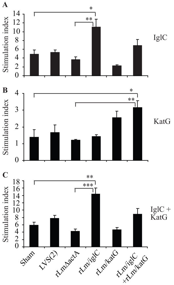
Figure 6.
Lymphocyte proliferation induced by intradermal immunization with attenuated L. monocytogenes vaccines expressing F. tularensis IglC or KatG. Groups of 3 BALB/c mice were sham-immunized, immunized i.d. twice with 1 × 10⁴ CFU LVS, or 1×10⁶ CFU recombinant L. monocytogenes vaccines expressing IglC (rLm/iglC), KatG (rLm/katG), or the combination of both at weeks 0 and 3. Six days after the second immunization, mice were euthanized and their splenocytes isolated and assayed for lymphocyte proliferation in response to recombinant IglC (A), KatG (B) or the IglC plus KatG (C) proteins. The mean CPM varied from group to group, ranging from 355 to 564 in the absence of the F. tularensis protein and from 493 to 4840 in the presence of the F. tularensis protein. The Stimulation Index (SI) was determined by dividing the mean CPM obtained in the presence of the F. tularensis protein by the mean CPM obtained in the absence of F. tularensis protein. Standard errors of the means of triplicate samples from three mice are shown. Asterisks indicate that the differences in SIs between the indicated groups are significant. *, P<0.05; **P<0.01; and ***P<0.001 by one-way ANOVA with Bonferroni’s post test.

