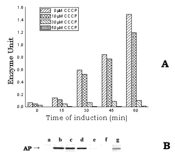
Figure 4.
A. Level of active AP in E. coli MPh42 cells grown in the presence of different concentrations of CCCP. Cells were initially grown to log phase (~1.5 × 10⁸ cells/ml) at 30°C in complete MOPS medium and were then transferred to phosphate-less MOPS medium. The re-suspended cells were divided in different parts to treat with the different concentrations of CCCP (0, 10, 30 and 50 μM). The divided cell cultures were then allowed to grow further at 30°C for induction of AP. At different intervals of time, a 1.0 ml cell aliquot was withdrawn from each culture to assay the active AP level. B. Western blot of the different fractions (periplasmic, cytoplasmic and membrane) of E. coli MPh42 cells grown in the presence of CCCP (50 μM). After allowing induction of AP for 30 min, the periplasmic, cytoplasmic and membrane fractions were isolated from equal number of each of the CCCP-treated and the control cells and the western blotting experiment was subsequently performed using anti-AP antibody. Lanes (a, b, c) and (e, f, g) represent the membrane, periplasmic and cytoplasmic fractions of control and CCCP-treated cells respectively; lane d represents purified AP.

