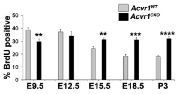
Figure 2.

Compared with wild-type, Acvr1CKO cells have reduced proliferation during early lens formation but increased proliferation at later stages. Sections of embryo heads or postnatal eyes were labeled for BrdU, and the percentages of BrdU-labeled nuclei in Acvr1WT and Acvr1CKO lens placodes and epithelia were counted from E9.5 to P3. ** P < 0.01; ***P < 0.001; ****P < 0.0001.
