Figure 5.

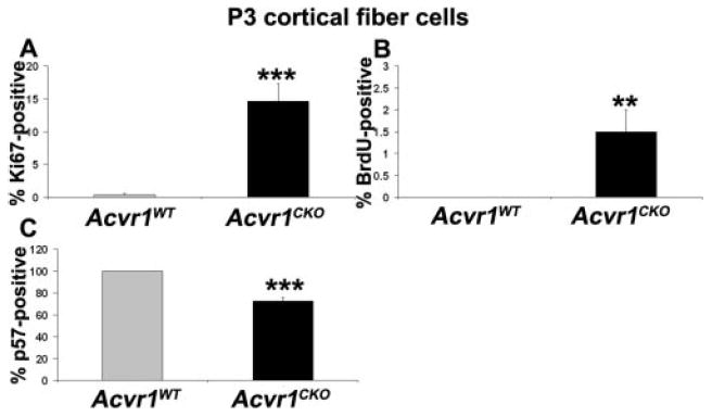
A subset of Acvr1CKO fiber cells fails to exit from the cell cycle. Sections of P3 Acvr1WT and Acvr1CKO eyes were labeled with antibodies against Ki67, BrdU, or p57, and the percentage of labeled nuclei in the cortical fiber cells was counted. There was a significant increase in the Ki67- (A) and BrdU-labeling index (B) and a significant decrease in the p57KIP2-labeling index (C) in Acvr1CKO cortical fiber cells compared with Acvr1WT fiber cells. **P < 0.01; ***P < 0.001.
