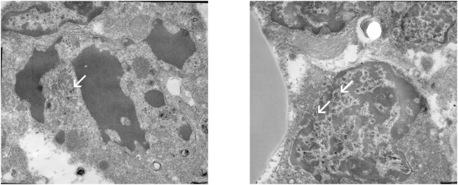
Figure 1. Electron micrographs showing viral particles in a sample of patient no. 4.
a) destroyed merkel cell carcinoma cell with viral particles (white arrow) measuring 50 nm, mingled with nuclear fragments and multiple smaller ribosomes (20 nm). b) penetration of virions through the nuclear membrane (white arrows) toward the cytoplasm (on the left). (original magnification; 12000×).

