
Figure 4.
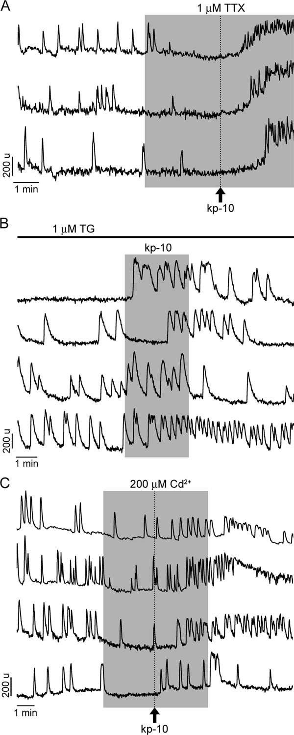
Identification of the source of calcium for kp-10-induced calcium signaling. A, A small fraction of cells (21%) with blocked spontaneous calcium oscillations by TTX responded to kp-10 with increase in [Ca2+]I, suggesting a role of intracellular calcium pools in response. Note the pattern of calcium signals in three representative cells. In the residual cells (79%) bathed in TTX-containing medium, kp-10 was completely ineffective, suggesting dependence of agonist action on the extracellular calcium pool. B, Consistent with the major role of calcium influx in kp-10 action, in 72% of cells with depleted ER calcium pool with TG, kp-10 also induced rise in [Ca2+]i. Four representative traces are shown. C, Only in a fraction of cells (44%, four representative traces are shown) bathed in medium contain cadmium, a kp-10-induced rise in [Ca2+]i occurred, further indicating the relevance of intracellular calcium pools for kp-10 action for a subpopulation of cells. In this and following figures, vertical dotted lines indicate the moment of kp-10 application, which was removed at the same time as cadmium.