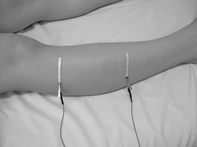
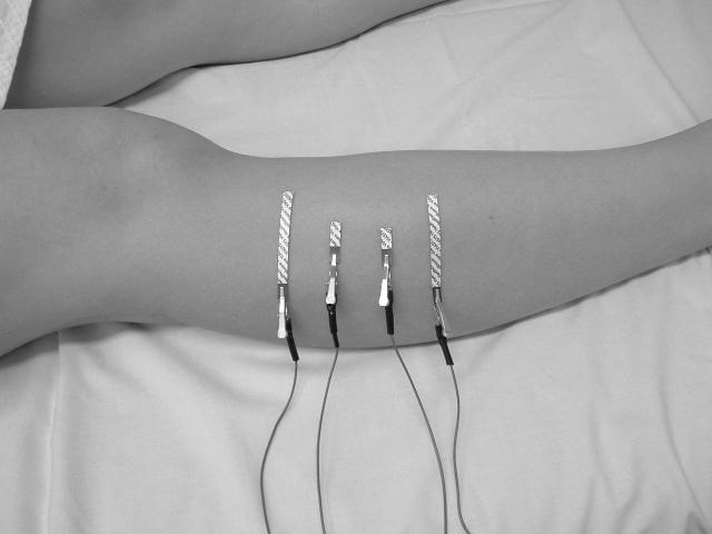
Figure 1.
Voltage electrode configurations for EIM using far- (Figure 1a) and near-electrode montages (Figure 1b). The far-electrode configuration for tibialis anterior uses current-injecting electrodes placed on the dorsal surfaces of the feet (not shown), at a distance from the site of voltage measurement. Current electrodes in the near-electrode montage are adjacent to the voltage-measuring electrodes, as shown in Figure 1b.


