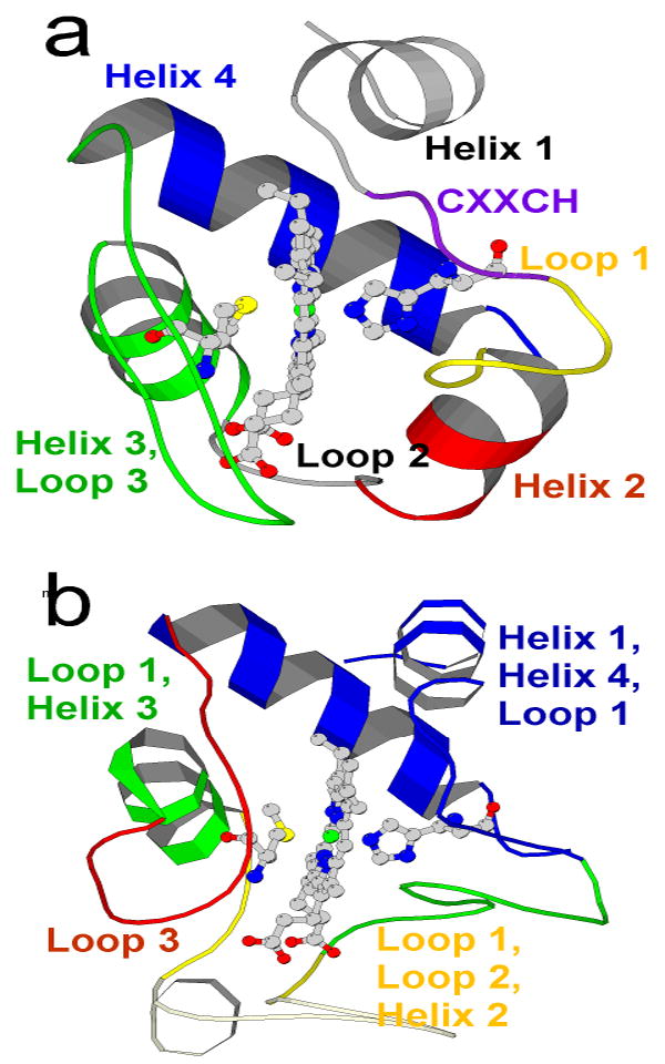
Fig. 2.

a The proposed foldons of Pa-cyt c; the extrapolated free energies ΔGHX,0 for the foldons are violet > blue > green > yellow > red. Helix 1 and loop 2 (grey) could not be placed into any of the foldons. b Foldons of h-cyt c [12, 26]. The lowest energy h-cyt c foldon is found within the omega loop at the bottom of the molecule as shown and is termed “nested yellow.”
