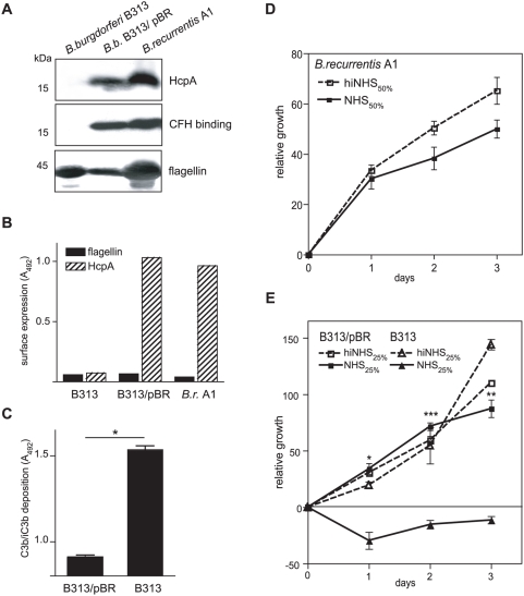
Figure 9. Ectopic expression of HcpA in serum-sensitive B. burgdorferi B313.
(A) Expression of HcpA by transformed B. burgdorferi B313 was assessed using immunoblot analysis. Whole cell lysates were separated by SDS-PAGE, transferred to nitrocellulose and probed with mAb BR-1 (upper panel) or analyzed for CFH binding by incubation with NHS and a CFH-specific mAb (JHD7, middle panel) followed by peroxidase conjugated IgG. For control, a flagellin-specific antibody (LA21) was used (lower panel). (B) Surface expression of HcpA as analyzed by whole cell ELISA using mAb BR-1. As control, a flagellin-specific mAb LA21 was employed. (C) C3b deposition on the surface of B. burgdorferi B313/pBR cells incubated with 10% NHS was determined using a whole cell ELISA as described above. Values represent the mean of triplicates±SEM. *, P<0.0001. To investigate serum susceptibility to human serum B. recurrentis A1 (D), B.burgdorferi B313 and transformed B. burgdorferi B313/pBR cells (E) were incubated with the indicated concentrations of NHS (dashed line) or heat-inactivated serum (solid line) at 30°C for 3 days. Cells were stained with a nucleic acid dye and the relative growth was determined by measurement of the fluorescence intensities. Values represent the mean±SEM of a single experiment performed in triplicate that is representative of three independent experiments. *, P<0.01; **, P<0.001; ***P<0.0001.

