Figure 6.
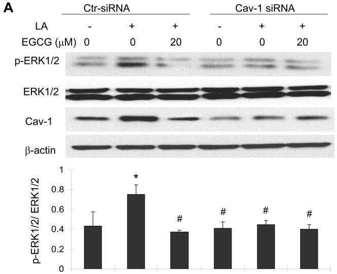
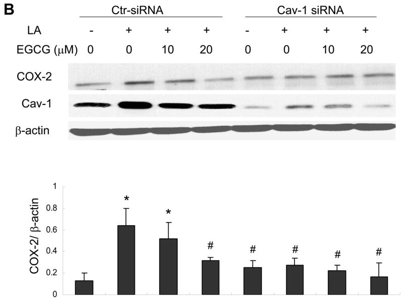
Caveolin-1 silencing mimics the protective effects of EGCG on linoleic acid-induced ERK1/2 phosphorylation (Figure 6A) and activation of COX-2 (Figure 6B). Endothelial cells were transfected with siRNA for caveolin-1 (Cav-1 siRNA) or with control siRNA (Ctr- siRNA) and treated with EGCG (20 μM) for 12 h, followed by exposure to linoleic acid (LA, 90 μM) for 10 min (Figure 6A) or 6 h (Figure 6B). Cell lysates were probed with caveolin-1 (Cav-1), COX-2 and β-actin antibodies or with anti-p-ERK1/2 and anti-ERK1/2. Protein expression was determined by western blot analysis. The western blot shown in each figure represents one of three experiments. Densitometry results shown in parallel represent the mean ± SEM of three independent experiments. *Significantly different compared to control cultures. #Significantly different compared to cultures treated only with LA (Ctr-siRNA).


