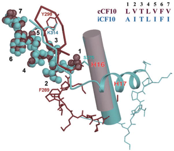
Fig. 6.
Comparison of the atomic interactions between amino acid residues of bound iCF10 and cCF10 with the PrgX C-terminus. cCF10 is shown as dark red van der Waals spheres; iCF10 is shown as light blue spheres. Positions of the side-chains of iCF10 and cCF10 are numbered 1–7. Dark red ribbon and sticks indicate the PrgX C-terminus in the cCF10 complex; light blue ribbon and sticks indicate the conformation in the iCF10 complex. Sequence of iCF10 and cCF10 are given in the upper right. The conserved helix 16 is shown as a cylinder.

