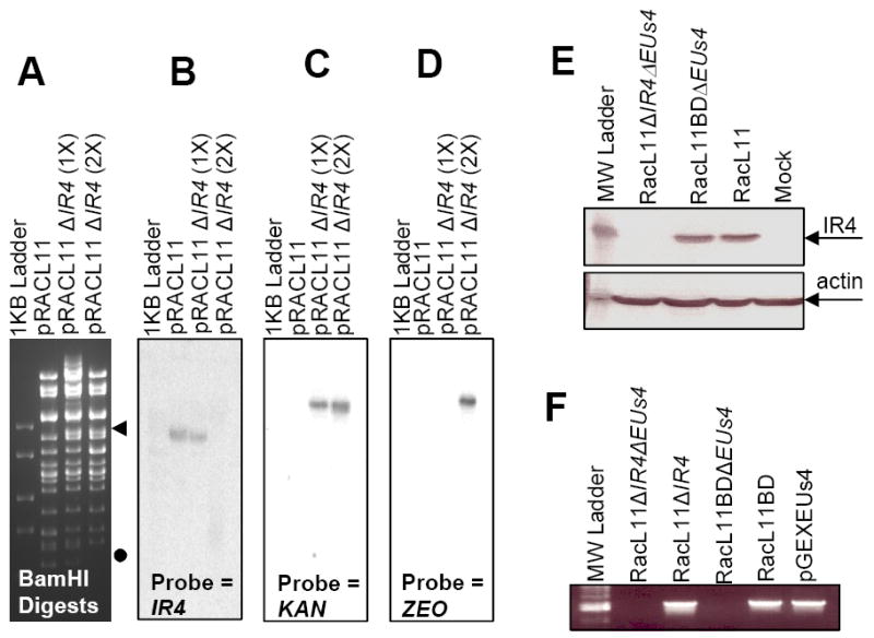
Fig. 1.

Confirmation of IR4 deletion mutant BAC. Panel A: BamHI restriction enzyme analysis of BAC DNA. ◀ = band added as result of IR4 deletion; ● = band removed as result of IR4 deletion. Panels B to D: Southern blot analyses of digested BAC DNAs. Panel E: western blot analysis of extracts of NBL-6 cells infected with EHV-1 wt (RacL11), EHV-1 RacL11 deleted of both the IR4 and EUs4 genes (RacL11ΔIR4ΔEUs4), or EHV-1 RacL11 deleted of EUs4 (RacL11ΔEUs4). Panel F: PCR amplification using primers specific for EUs4 of viral DNA lacking both IR4 and EUs4 genes (RacL11ΔIR4ΔEUs4), viral DNA lacking IR4 with the EUs4 gene restored (RacL11ΔIR4), viral DNA lacking EUs4 gene (RacL11ΔEUs4), viral DNA with the EUs4 gene restored (RacL11BD), and a control plasmid harboring the EUs4 ORF (pGEXEUs4). Methods for restriction enzyme digestion, Southern blot analysis, and PCR protocols are described in the Materials and Methods.
