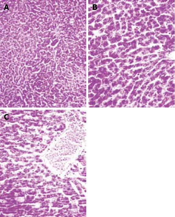
Figure 4.

Hematoxylin and eosin-stained sections of rat liver. A: Sham-operated group. B: I/R group, vacuolization was frequent in the 45 min ischemia-reperfused tissues. Irregularity, pale (atypical) nuclei, disintegrated cytoplasm, and infiltration of leukocytes were seen. C: Similar changes were observed in the 60 min ischemia-reperfused group (× 400).
