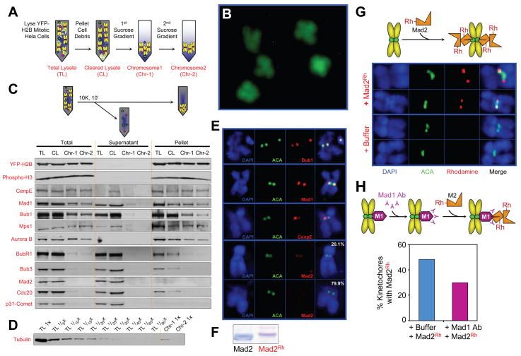
Fig. 1. Unattached kinetochores on purified chromosomes recruit Mad2.
(A) Schematic of chromosome purification from mitotic HeLa cells stably expressing YFP-H2B histone. Cells were collected after 16 hours in colcemid, lysed, cell debris removed by pelleting and the chromosome containing supernatant was fractionated on sequential sucrose gradients. (B) Morphology of purified chromosomes detected by fluorescence of YFP-H2B on coverslips without fixation. (C) Protein constituents of purified chromosomes assessed by immunoblotting after pelleting. (D) Tubulin levels remaining in purified chromosomes, along with a dilution series of the initial cellular input. (E) Indirect immunofluorescence for detection of Mad1, Bub1, CENP-E and Mad2 on isolated chromosomes. (Blue) Chromosomes stained with DAPI; (Green) Anticentromere (ACA) antibodies; (right panel) merged image. (F) Purified recombinant Mad2 before and after covalently labeling with rhodamine, assessed by Coomassie staining. (G) Purified chromosomes were incubated with rhodamine-labeled Mad2, fixed, stained for (blue) DAPI and (green) ACA, and imaged by deconvolution microscopy. (H) Chromosomes were incubated for 10 min with anti-Mad1 antibody, then with rhodamine-labeled Mad2, and finally fixed, stained, imaged as in (G), and scored for Mad2 localization.

