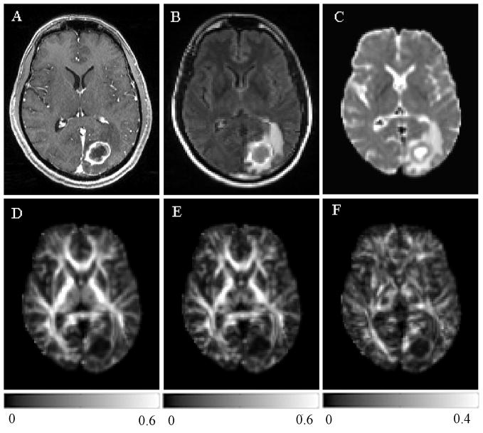
Figure 3.
A 56 year old male with metastatic lung adenocarcinoma in the left occipital lobe. There was no hemorrhage based on T1 and T2-weighted images (not shown). Transverse contrast-enhanced T1-weighted (A) and FLAIR (B) images show a ring-enhancing lesion with extensive edema, similar in appearance to the glioblastoma shown in Figure 2 (A and B). ADC map (C) shows restricted diffusion of the enhancing part. Lower FA (D), CL (E) and CP (F) are noticed from the enhancing part relative to normal appearing white matter compared with the glioblastoma.

