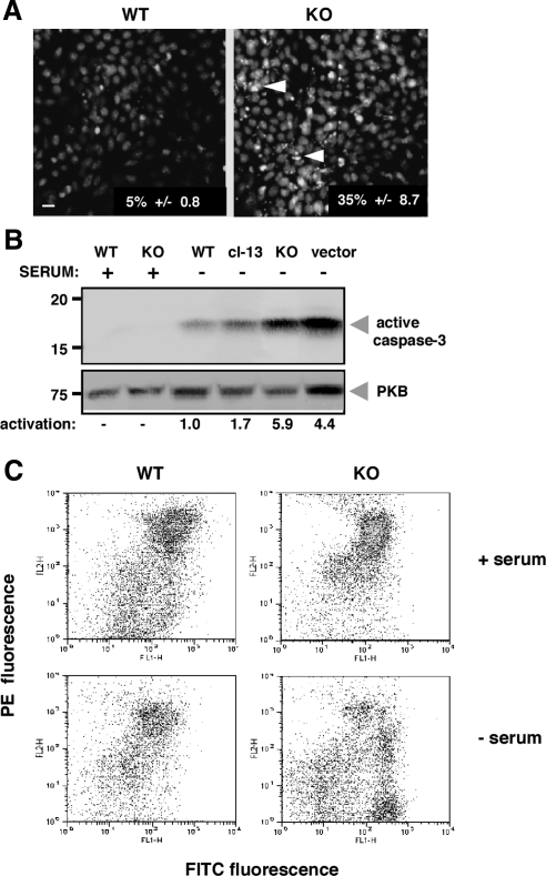
Figure 2.
grp94−/− cells undergo apoptosis in the absence of serum. (A) WT and grp94−/− cells (KO) were incubated for 12 h in the absence of serum and then stained with Hoechst 33258 (bis-benzidine). The percentages of cells with condensed chromatin (arrowheads) were derived from counting at least 250 cells each, and the numbers listed in the figure represent the mean ± SD of three independent experiments. Scale bar, 20 μm. (B) To measure activation of caspase 3, cell extracts (40 μg) were derived from either WT, grp94−/− (KO) cells, or KO cells transfected with a grp94 expression vector (cl-13) or with an empty vector (cl-3). Lysates were separated by SDS-PAGE and immunoblotted with an antibody specific for cleaved caspase-3 (17-kDa band). The level of the cytosolic protein kinase B (PKB) served as a loading control. The results from densitometric analysis of the autoradiograms are shown below the gel, expressed in relative units of activated caspase 3. (C) FACS analysis of JC-1–stained cells. WT or grp94−/− cells were loaded with JC-1, a mitochondrial voltage-sensitive dye and analyzed by FACS after incubation in normal or serum-free media. Live cells are mostly orange (FL-2 axis), and the green shift (FL-1 axis) in serum-free condition reflects cells with depolarized mitochondria undergoing apoptosis.

