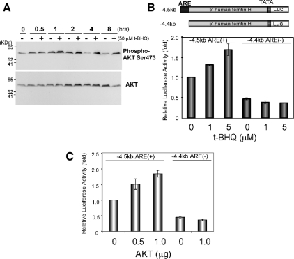
Figure 2.
Ferritin H ARE is activated by t-BHQ treatment or AKT in Jurkat cells. (A) Growing Jurkat cells were either untreated (−) or treated with 50 μM t-BHQ (+), and Western blot was carried out for phospho-Ser473 AKT (top) and AKT (bottom). Representative results from four independent experiments are shown. (B) Jurkat cells (1–2 × 10⁷) were transfected with 1 μg of −4.5kbARE(+)- or −4.4kbARE(−)-ferritin H luciferase and then treated with 1 or 5 μM t-BHQ for 24 h, or (C) along with 0.5 or 1 μg of pcDNA3.1T7-AKT1 transfection. Luciferase expression from −4.5kbARE(+)-luciferase without t-BHQ treatment in B, or with 1 μg of pcDNA3.1T7 empty vector in C was set to 1.0. Means ± SEs from three independent experiments are shown.

