Figure 3.
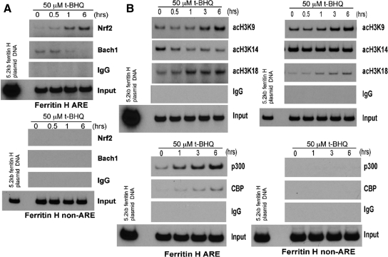
Alterations in histone H3 acetylation along with recruitment of Nrf2 and p300/CBP to the ferritin H ARE in t-BHQ treated Jurkat cells. (A) Jurkat cells (1–2 × 10⁶) were treated with 50 μM t-BHQ and subjected to chromatin immunoprecipitation with rabbit immunoglobulin G (IgG), anti-Nrf2 or anti-Bach1 antibody as described in Materials and Methods. The immunoprecipitates were subjected to semiquantitative PCR by using the ferritin H ARE and non-ARE primer sets. 0.5 pg of −5.2kb ferritin H-luciferase plasmid DNA was used as a template of the PCR-positive control and size marker of the PCR-amplified 0.15-kb (ARE) or 0.2-kb (non-ARE) DNA. Nonimmunoprecipitated DNA was also PCR amplified to assess the amount of input DNA for the ChIP assay. A representative of three independent experiments is shown. (B) Jurkat cells (1–2 × 10⁶) treated with 50 μM t-BHQ were similarly subjected to ChIP assays with rabbit IgG or antibodies against acetyl histone H3-K9, -K14, -K18, p300, and CBP. A representative of three independent experiments is shown.

