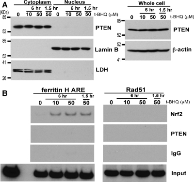
Figure 7.
Assessment of Nuclear PTEN and its association with the ferritin H ARE in K562 cells. (A) K562 cells (3 × 10⁶) were treated with 0, 10, or 50 μM t-BHQ for 1.5 or 6 h and subjected to isolation of whole cell lysate, cytoplasmic, and nuclear fractions. Samples (50 μg) were analyzed for protein expression by Western blotting with anti-PTEN, anti-Lamin B, anti-LDH antibodies. (B) K562 cell (1 × 10⁶) were treated with 0, 10, or 50 μM t-BHQ for 1.5 or 6 h, and ChIP assays were performed using control IgG, anti-Nrf2, or anti-PTEN antibody. The immunoprecipitates were subjected to semiquantitative PCR by using primer sets for the ferritin H ARE or the PTEN-regulated Rad51 promoter (Shen et al., 2007).

