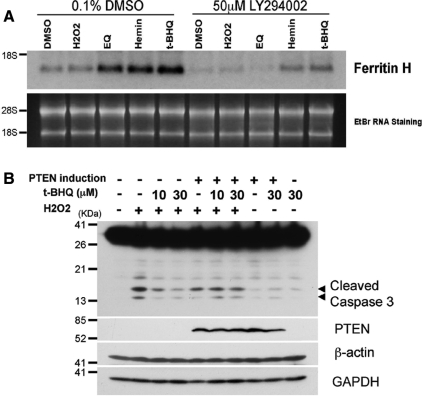
Figure 8.
Effect of PTEN expression on t-BHQ–mediated cytoprotection from hydrogen peroxide-induced apoptosis. (A) Jurkat cells (2 × 10⁶) were pretreated with 0.1% DMSO or 50 μM LY294002 for 1 h, followed by treatment with 0.25% DMSO, 25 μM H2O2, 50 μM ethoxyquin (EQ), 20 μM hemin, or 10 μM t-BHQ for 16 h. Total RNA was isolated and 5 μg each of RNA was subjected to Northern blotting analysis with human ferritin H cDNA probe. RNA staining with ethidium bromide is shown for comparative loading of RNA. Representative results from three independent experiments are shown. (B) PIJ17 cells (5 × 10⁶) were treated with 1 μg/ml doxycycline for 24 h, followed by pretreatment with 10 or 30 μM t-BHQ for 48 h before 100 μM hydrogen peroxide challenge for 6 h. We analyzed 50 μg of whole cell lysates by Western blotting using anti-caspase-3, anti-PTEN, anti-glyceraldehyde-3-phosphate dehydrogenase (GAPDH), or anti-β-actin antibody.

