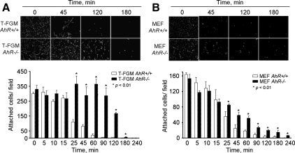
Figure 6.
AhR expression decreases cell adhesion to the substratum. (A) T-FGM AhR+/+ and T-FGM AhR−/− cells were seeded at the same cell density on plastic dishes and then induced to detach by using treatments with EGTA for the indicated times. Nuclei were stained with DAPI, and attached cells counted using the ImageJ software. (B) MEFs of the indicated genotypes were cultured at the same initial cell density and processed and analyzed as described in A. Data are shown as mean ± SEM for measurements made in duplicate in at least two independent cultures for each genotype and cell type. The p values for statistical comparison between genotypes are indicated.

