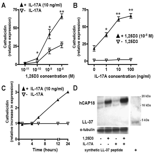
FIGURE 2.
IL-17A enhances the effect of 1,25D3 on cathelicidin mRNA and cathelicidin peptide hCAP18 in primary human keratinocytes. A, NHEK were treated with IL-17A (10 ng/ml) in the presence of increasing concentrations of 1,25D3 (10 −11–10−8 M) for 24 h. Cathelicidin transcript abundance was analyzed by quantitative real-time PCR. B, NHEK were treated for 24 h with 1,25D3 (10−8 M), and increasing concentrations of IL-17A (1–100 ng/ml). Cathelicidin was measured by quantitative real-time PCR. C, Keratinocytes were stimulated with IL-17A (10 ng/ml) in the presence of 1,25D3 (10−8 M). Cathelicidin induction as measured by quantitative real-time PCR at the indicated time points was calculated relative to the induction by 1,25D3 alone. Data are mean ±SD of a single experiment performed in triplicate and are representative of three independent experiments.*, p < 0.05 and **, p < 0.01, determined by Student’s t test. D, To evaluate cathelicidin peptide induction, NHEK were treated for 24 h with 1,25D3 (10−8 M), IL-17A (100 ng/ml), or the combination. Cathelicidin hCAP18/LL-37 protein expression was analyzed in NHEK lysates by Western blot using an Ab that detects hCAP18 and LL-37. Synthetic LL-37 peptide was used as a positive control (far right lane).

