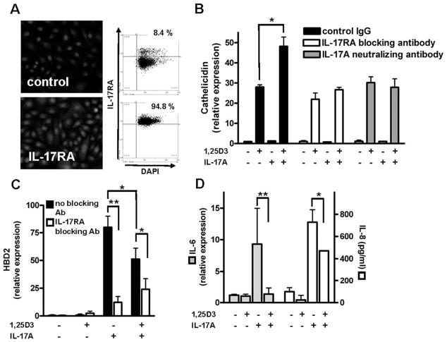
FIGURE 3.
NHEK express IL-17RA while 1,25D3 differentially affects IL-17A-induced gene expression in keratinocytes. A, Expression of the IL-17RA was confirmed in primary NHEK by immunofluorescence staining and TissueFAXS analyses. Cell nuclei were detected by DAPI. B, NHEK were stimulated with IL-17A (10 ng/ml) and 1,25D3 (10−8 M) for 24 h, and cathelicidin transcript abundance was measured by quantitative real-time PCR. In addition, cells were preincubated 2 h with an IL-17RA blocking Ab before stimulation. As an additional control, an IL-17A neutralizing Ab was added to culture medium containing IL-17A 2 h before the medium was applied to the cells. The 1,25D3 stimulation enabled IL-17A to increase cathelicidin, which was inhibited by the IL-17RA blocking Ab or the IL-17A neutralizing Ab. To evaluate the expression of other IL-17A regulated genes, NHEK were stimulated with IL-17A (10 ng/ml) and 1,25D3 (10−8 M) for 24 h and HBD2 (C) and IL-6 (D) transcript abundance was measured by quantitative real-time PCR. IL-8 expression (D) in cell culture supernatants was analyzed by ELISA. Cells were incubated with an IL-17RA blocking Ab 2 h before stimulation. HBD2 expression was induced by IL-17A alone, and induction was attenuated in the presence of 1,25D3. C, Further inhibition of IL-17A signaling inhibited HBD2 induction. D, Similar to HBD2 expression, IL-17A induced IL-6 (left) and IL-8 (right) expression that was decreased when 1,25D3 was present. Data are mean ± SD of a single experiment performed in triplicate and are representative of three independent experiments.*, p < 0.05 and **, p < 0.01, determined by Student’s t test.

