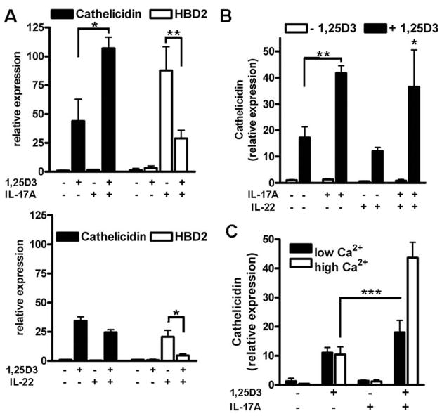
FIGURE 4.
Th17 cytokines IL-17A and IL-22 differentially affect antimicrobial peptide expression. To compare the effects of the Th17 cytokines, IL-17A (top) and IL-22 (bottom) keratinocytes were treated with IL-17A (10 ng/ml) or IL-22 (10 ng/ml) in the presence of 1,25D3 (10−8 M). Cells were harvested 24 h after stimulation and mRNA levels for cathelicidin and HBD2 were analyzed by quantitative real-time PCR. A, 1,25D3 enabled IL-17A to increase cathelicidin, whereas IL-22 had no effect. Both, IL-17A and IL-22 induced HBD2 in keratinocytes as measured by quantitative real-time PCR and again 1,25D3 attenuated HBD2 induction. B, The combination of IL-17A and IL-22 showed the same effect on cathelicidin transcript abundance as IL-17A alone. C, To evaluate the influence of cell differentiation on cathelicidin induction, NHEK were grown in medium containing low (0.06 mM) or high (1.7 mM) concentrations of calcium for 24 h. Cells were then stimulated with IL-17A (10 ng/ml) in the presence or absence of 1,25D3 (10−8 M) for another 24 h. Cathelicidin transcript abundance was analyzed by quantitative real-time PCR. Induction of cathelicidin by 1,25D3 did not change with increasing calcium concentration, but the effect of IL-17A was significantly stronger in keratinocytes treated with 1.7 mM calcium. Data are mean ± SD of a single experiment performed in triplicate and are representative of three independent experiments.*, p < 0.05; **, p < 0.01; and ***, p < 0.001, determined using Student’s t test.

