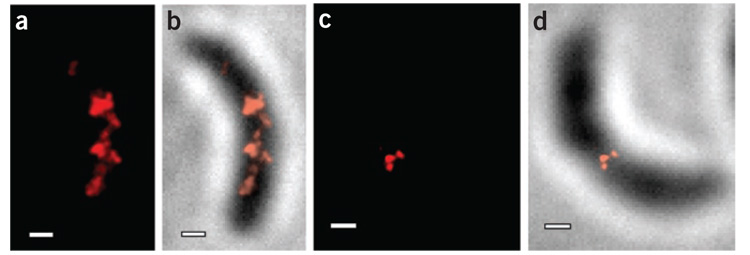
Figure 3.
TL-PALM images of EYFP-MreB in C. crescentus cells showing fewer punctuate spots than continuous-acquisition PALM. (a,b) Quasi-helical structure in a stalked cell. (c,d) Midplane ring in a predivisional cell. Fluorescence PALM images are shown in a and c. The PALM images in b and d are the same cells as in a and c, respectively, overlaid on a reversed-contrast white-light transmission image of the cell. Scale bars, 300 nm.

